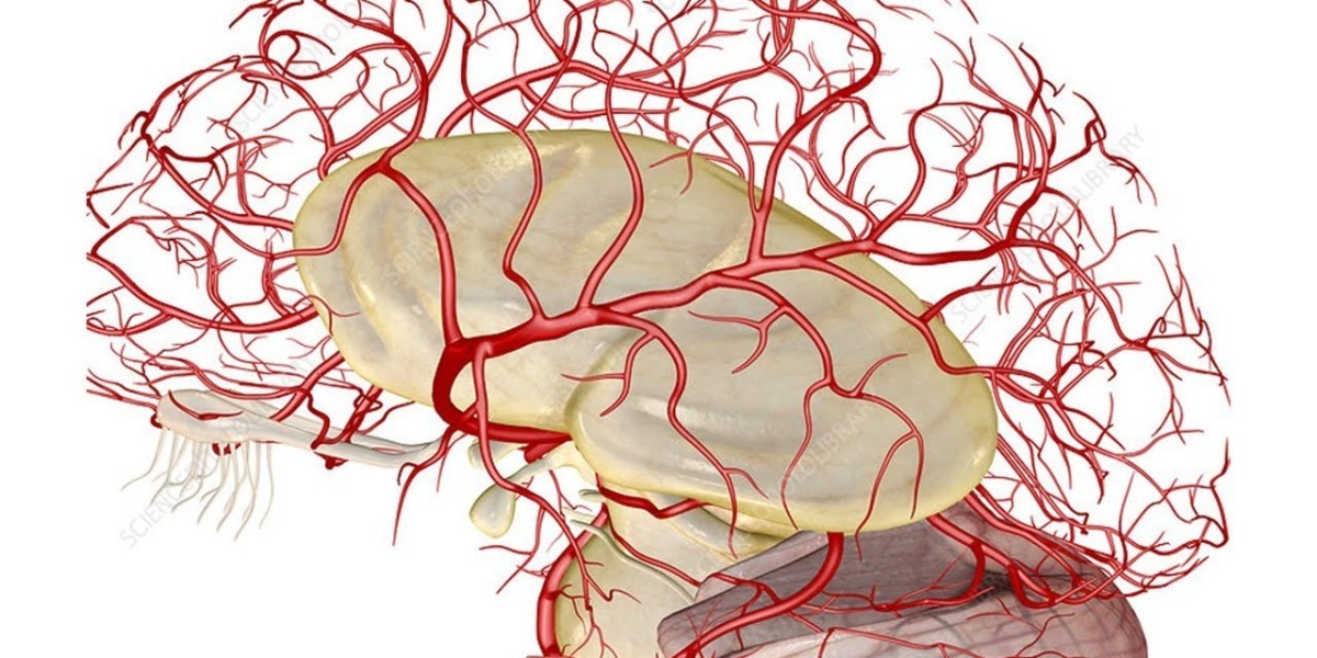
Subtraction angiography is a medical imaging technique used to assess the arteries in the brain and neck. During the procedure, a catheter is inserted into an artery, usually in the groin, and threaded through to the arteries supplying the brain. A contrast dye is then injected through the catheter and multiple X-ray images are taken to visualize the arteries. This allows doctors to evaluate for conditions like aneurysms, arterial narrowing (stenosis), blood clots, tumors and other vascular abnormalities.
The Procedure
Before the test, the patient is asked not to eat or drink anything for a few hours prior. On the day of the procedure, the patient lies flat on their back on an X-ray table. The skin over the groin or arm is cleaned and numbed with a local anesthetic. A thin tube called a catheter is inserted into the femoral or radial artery and guided through these arteries using real-time X-ray imaging until the tip reaches the arteries in the neck.
Once in place, a contrast dye is injected through the catheter into the arteries. As the dye circulates through the brain vasculature, a series of very quick X-ray images called angiograms are taken at intervals. The contrast dye allows the arteries to be seen clearly on these images. The entire procedure takes about 45-60 minutes. After the catheter is removed, pressure is applied to the puncture site to prevent bleeding. The patient is monitored for a few hours before being discharged.
Potential Risks
Though Cerebral Angiography is a low risk procedure, there are some potential complications:
- Bleeding or bruising at the puncture site: This is the most common side effect. Applying pressure can prevent this.
- Reaction to contrast dye: Some people may get an allergic-like reaction like flushing, hives or breathing difficulties. This is treated with medication.
- Contrast-induced nephropathy: The dye can potentially harm kidney function but the risk is low. Adequate hydration before and after helps prevent this.
- Stroke: Getting a blood clot or bubble of air in the brain during catheter manipulation is very rare but possible. The risk is less than 0.1%.
- Artery damage: There is a very small chance of puncturing or tearing the arteries during catheter insertion.
- Irregular heartbeat: Some may experience changes in heart rhythm due to dye or anxiety during the test.
Precautions are taken to minimize these risks. Subtraction angiography is still considered very safe when performed by experienced radiologists in high volume centers.
Uses of Cerebral Angiography
Some common conditions where subtraction angiography helps in diagnosis and management include:
Aneurysms: It can precisely locate brain aneurysms, determine their size and shape. This assists in planning treatment like surgery or coil embolization.
Arteriovenous malformations (AVM): These abnormal cluster of blood vessels in the brain appear clearly on angiograms, helping confirm diagnosis. It also maps their extent and supply.
Stroke: When the cause of stroke is uncertain, angiography rules out blood vessel blockage or aneurysm as the cause and identifies other vascular issues.
Tumors: Certain brain tumors like meningiomas involve blood vessels which can be seen. It provides info on tumor vascularity before surgery.
Abnormal arteries: Congenital defects, arterial dissections, stenosis etc affecting cerebral blood flow are detectable.
Headaches: Patients with puzzling headaches and normal CT/MRI scans occasionally have vascular abnormalities found on angiograms.
Post treatment assessment: It helps evaluate the results and efficacy of earlier surgeries, coiling or other interventional procedures for conditions like AVMs.
Subtraction angiography remains an invaluable tool that provides unparalleled details of the intracranial blood vessels when less invasive imaging leaves questions unanswered. This forms the basis for effective diagnosis and management planning by neuroangiographers and neurosurgeons.
For More Insights Discover the Report In language that Resonates with you
Get more insights: Cerebral Angiography
Explore more Article: Immunotherapy Drugs Market
About Author:
Money Singh is a seasoned content writer with over four years of experience in the market research sector. Her expertise spans various industries, including food and beverages, biotechnology, chemical and materials, defense and aerospace, consumer goods, etc. (https://www.linkedin.com/in/money-singh-590844163)


